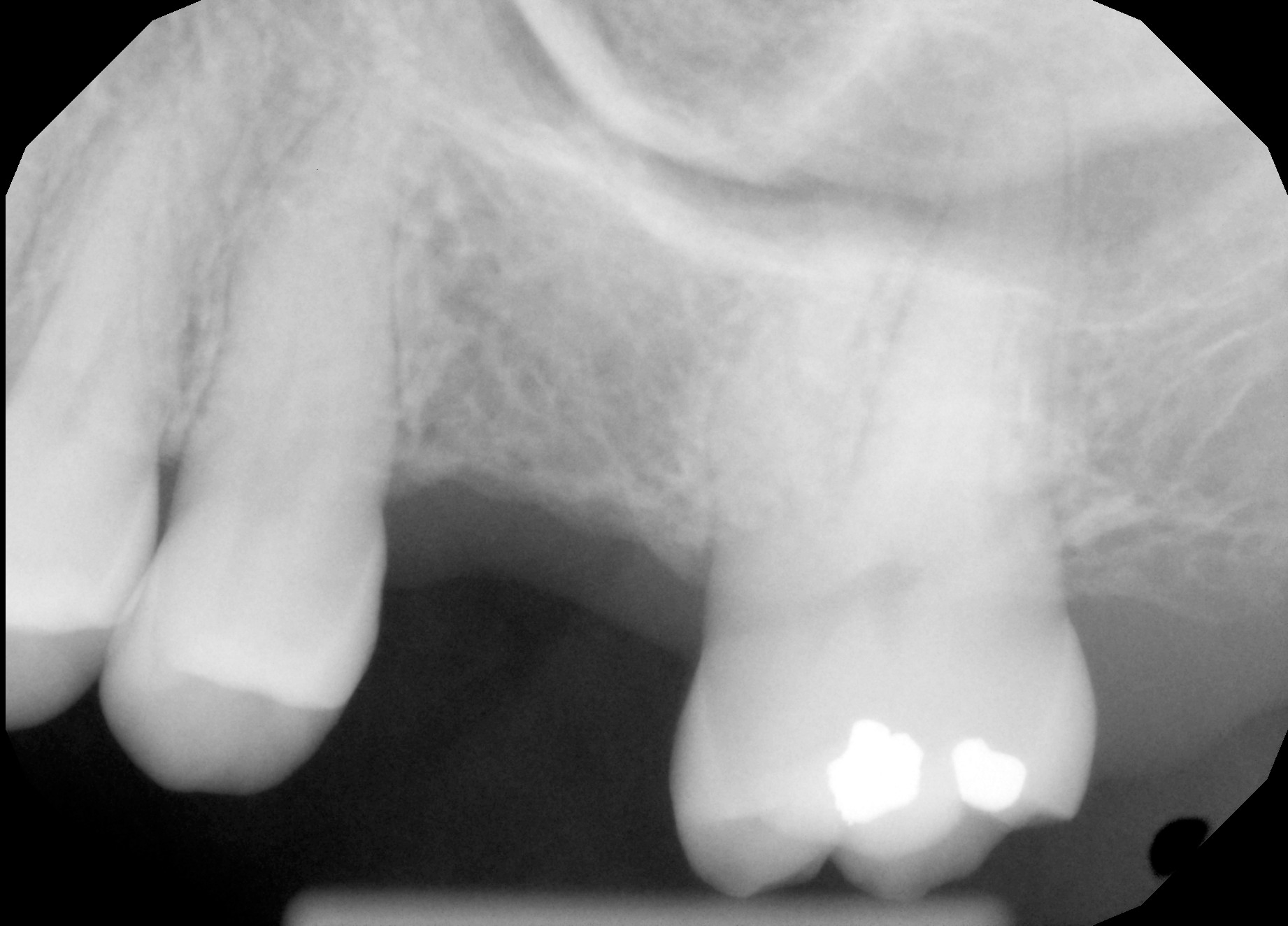
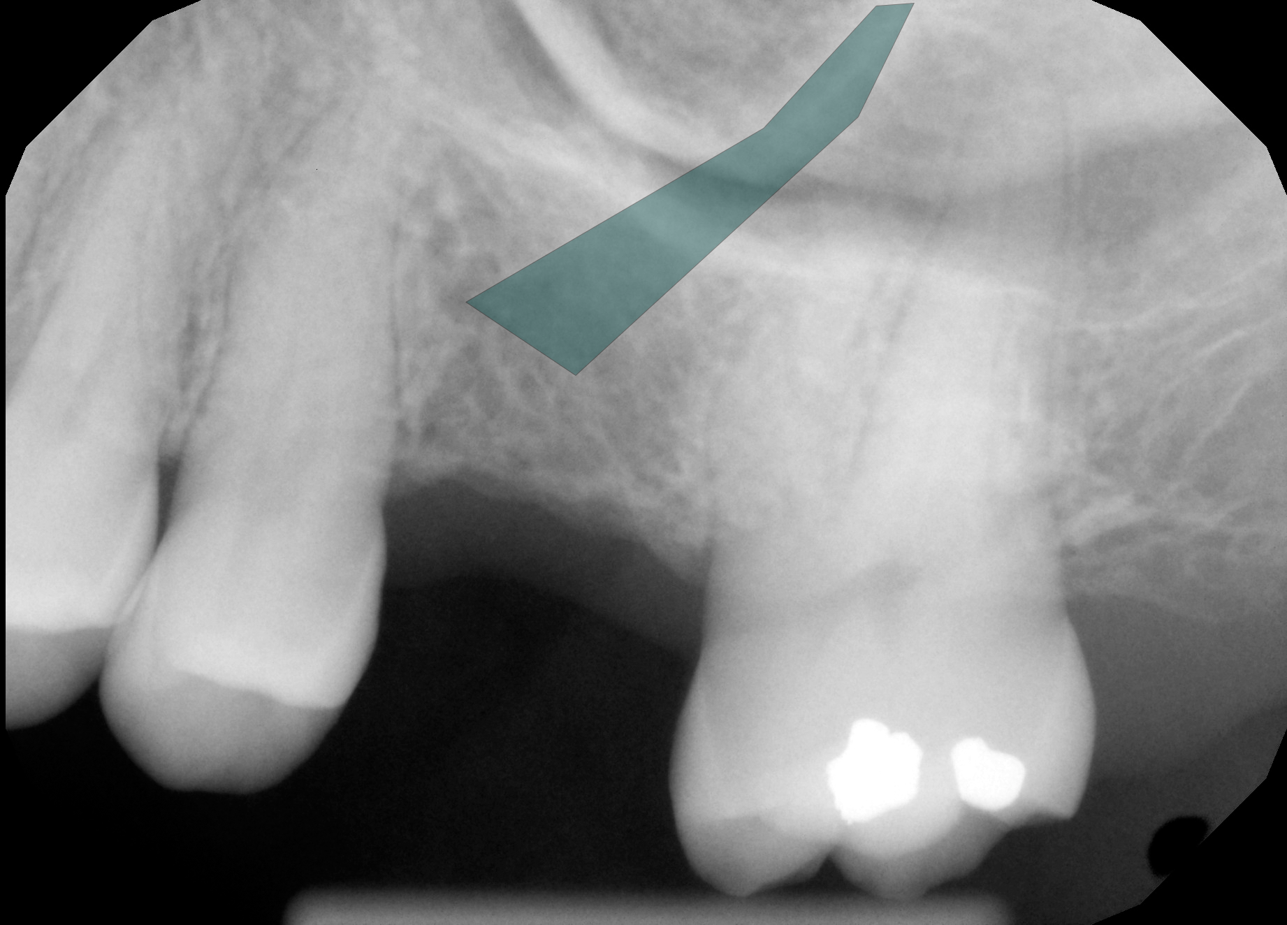
Radiographic Analysis (X-Ray)
Forensic identification of anomalous shape in extraction cavity.
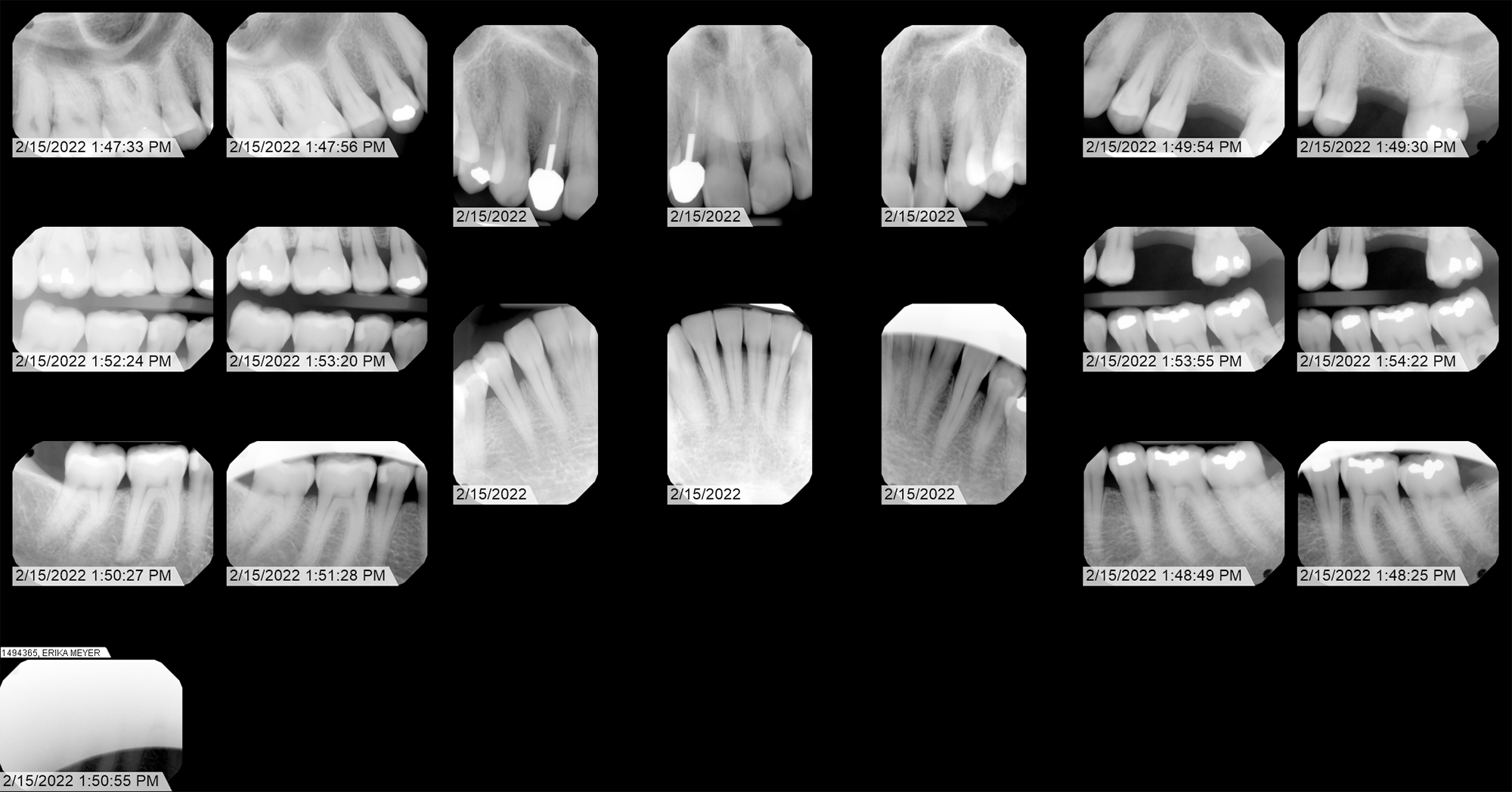
Technical Metadata (DICOM)
| Source Format | DICOM (Medical Standard) |
| Export Method | Lossless 16-bit PNG |
| Sensor Type | Intraoral Digital Sensor |
| Capture Date | Feb 15, 2022 | 13:49:30 |
Case Narrative: Tooth 14
This imaging is forensic support for my personal experiences related to pain, inflammation, swelling, heating, and vibrating sensations in this area and surrounding tissues, and it also supports results of other tests, in particular bug detector detection of HF signals from specific areas and types of dentistry, including from tooth 14, when it was intact and covered by a crown.
Tooth 14 was an otherwise healthy tooth which suddenly developed sensitivity in late spring of 2014. A dental visit determined that the tooth was cracked and unrecoverable. A root canal was performed and a crown placed over the tooth. I was not asked what type of crown I wanted: Willamette Dental selected a PFM (porcelain fused to metal) crown. The crown was over-large, and was never comfortable. In 2017 I picked up signals from the crown with my Aceco FC6002MKII bug detector. After I called Willamette Dental to report and complain about this, I no longer picked up signals from the crown. However, beginning in 2019 (after the bug detector had been stolen and sabotaged) the crown became increasingly problematic with effects such as heating, vibrating - things that to me were clearly electrical attacks focused at the crown and surrounding tissues of my head and neck. I was told that the crown could not be replaced, and that my only option was to leave it intact, or pull the tooth. On January 10, 2020, the tooth was pulled by Dr. Ashish Patel of Head & Neck Associates.
This featured x-ray, in my opinion, clearly shows an anomalous artifact embedded in my jawbone. This would have had to have been placed into the extraction cavity after the extraction and prior to suture. Dr. Patel is the only one who could have inserted this object - which appears to be a bioelectronic transceiver - into my jaw. Over time, the cavity fills with bone tissue, so the implant is now embedded in the jawbone.The significance of this radiograph is further reinforced by a history of institutional resistance encountered during attempts at verification. Despite significant effort throughout 2022, I was unable to obtain original DICOM files from either Advantage Dental or Roots Dental. However, in 2026 I was able to locate and cite the federal and state laws requiring them to provide me with that information: HIPAA (45 CFR § 164.524) and Oregon Administrative Rule (OAR) 818-012-0030. As of this writing Advantage Dental has provided me with those files, and to their credit, they did so quickly. Willamette Dental has also provided DICOM files, giving historical perspective. Roots dental did not provide DICOM or original files - and I believe there is a reason for this (to be continued...)
Under the principles established by the 21st Century Cures Act and SWGDE forensic standards, original DICOM data is considered the "Primary Evidence." Prior to this year, both Advantage Dental and Roots Dental insisted on substituting lower-resolution exports, effectively preventing the authentication of sensor-level metadata.
Last updated March 28, 2026
Secondary Exhibits
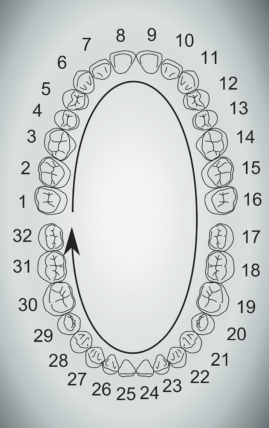
Dental map showing numbered teeth. Tooth 14 extracted Jan 10, 2020